HIFU Treatment for Adenomyosis | Aegis Roen HIFU Center




Main menu

Section menu
Page title
depends on advanced HIFU skill and precise analysis.
Page content
Adenomyosis When the uterine lining or similar tissue grows outside the uterine cavity (uterine muscle, ovaries, fallopian tubes, peritoneum, ligaments around the uterus, bladder, vaginal wall, etc.), it is called endometriosis, and when it grows inside the uterine muscle, it is called adenomyosis. It is reported to be most common in women in their 40s and 50s.
and severe pain, so it
so it needs to be treated.
Adenomyosis is when tissue from the uterine lining grows into the uterine wall. It is considered a type of endometriosis.
It also causes severe period pain, infertility, heavy periods, irregular bleeding and pelvic pain.
in young unmarried women in their 20s? Statistics show that more young women in their 20s and 30s have uterine conditions such as fibroids, adenomyosis and endometriosis. Rates are rising fast in women in their 20s and 30s. Many are not aware of uterine disease, skip regular check-ups, and dismiss severe period pain or back pain as ordinary PMS, missing the right time for treatment.
| 20s | 30s | 40s | 50s | 60s |
|---|---|---|---|---|
| 9,090 women | 56,630 | 144,489 | 77,568 | 12,315 |
whose lesions have no clear border, depends on advanced HIFU skilland precise analysis.
Unlike fibroids, adenomyosis does not form distinct nodules, so it has no clear border. It is usually about as small as a fibroid, and often occurs together with fibroids.
-
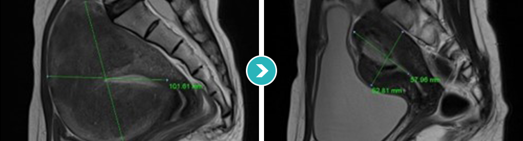 Diffuse type
DIFFUSE TYPE ADENOMYOSIS
Diffuse type
DIFFUSE TYPE ADENOMYOSIS
-
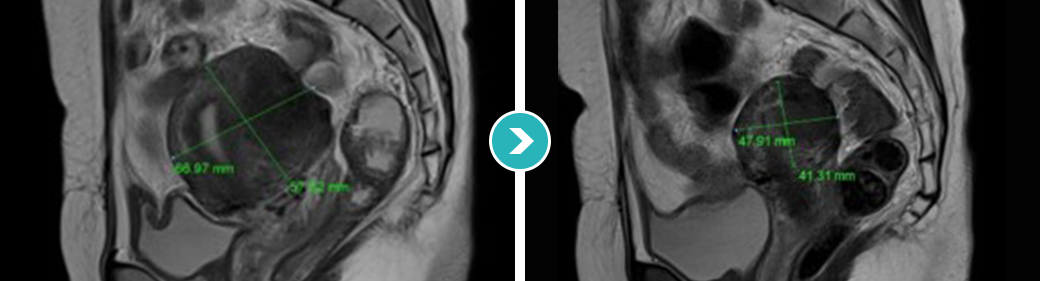 Nodular type
NODULAR TYPE ADENOMYOSIS
Nodular type
NODULAR TYPE ADENOMYOSIS
-
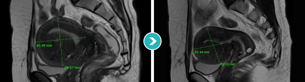 Sclerotic type
SCLEROTIC TYPE ADENOMYOSIS
Sclerotic type
SCLEROTIC TYPE ADENOMYOSIS
-
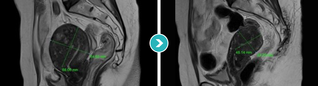 Cystic type
CYSTIC TYPE ADENOMYOSIS
Cystic type
CYSTIC TYPE ADENOMYOSIS
The organs in the body sit close together,
so after HIFU for adenomyosis
the doctor must make sure the lesion was well treated with ultrasound heat
from the inside to just inside its outer edge, and that
the heat did not affect nearby organs or nerves.
This requires a specialist with advanced
HIFU skill.
Future pregnancy and childbirth
may become impossible. If an unmarried woman's gynecologic ultrasound shows fibroids or another uterine condition, she should decide on treatment with a specialist. Treating it with surgery such as removal of the fibroid or the uterus can make future pregnancy and childbirth impossible.
Surgery that removes the whole uterus because the fibroid is large
Surgery that removes the lesion with instruments through small cuts instead of a large abdominal incision
A treatment that uses the heat created when high-intensity ultrasound energy is focused on one point to destroy the tissue
Early adenomyosis treatment system
The Roen AI-assisted HIFU program preserves the uterus, is safer than surgery, keeps pregnancy and childbirth possible, and involves no incision or bleeding
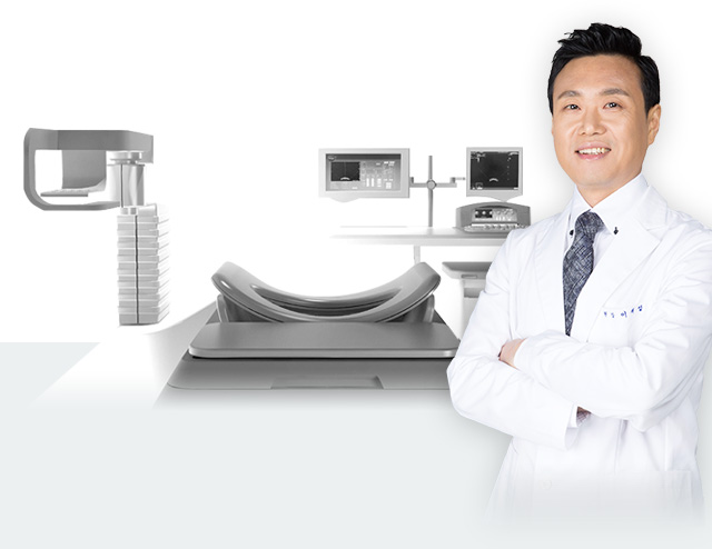
Artificial Intelligence HIFU System
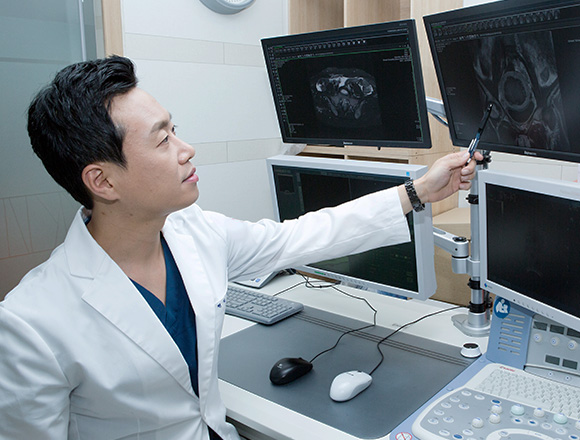
High-resolution MRI images and real-time ultrasound images are combined into a 3D view that tracks the uterus and tumor as they move with the patient's breathing and body movement, so that high-intensity focused ultrasound energy treats them safely.
Artificial Intelligence Check up System
For accurate diagnosis of tumors in the uterus, and to follow every possible change during recovery after HIFU, we run a specialized uterine check-up program for ongoing follow-up.
interface
The best treatment for humansMapping the path of the ultrasound energy for safe treatment. The key to HIFU is delivering focused ultrasound energy precisely to the lesion. Precise image analysis shows both where the lesion is and the path the ultrasound energy will take.
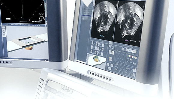
Advanced image fusion technology
Artificial Intelligence - Software SystemThe body is always moving. Accurate treatment requires tracking the lesion's position in real time as it moves. This is what makes treatment at Aegis Roen OB-GYN safer.

for safe treatment
Precise PositioningWe set the exact entry path of the high-intensity focused ultrasound energy to match the patient's body shape and the position and size of the uterus and tumor.

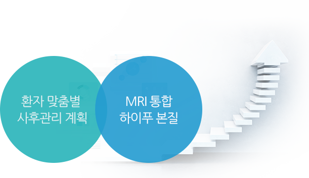
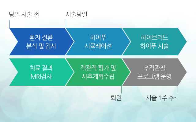
3STEP HIFU PROGRAM Since 2010, an MRI-integrated HIFU program at every step, dedicated to the safe treatment of fibroids and adenomyosis
MRI before treatment to check whether a lesion could become malignant and to look for hidden malignant lesions
Ultrasound and MRI monitoring during treatment
Results review and objective evaluation after HIFU
=
Non-surgical treatment system for fibroids ∙ adenomyosis
MRI is used to analyze the fibroid (location, number, size, shape, etc.), its blood supply, nearby organs and structures, and a clear path for the ultrasound. Detailed ultrasound then analyzes the number of fibroids to treat, the number of treatment slices, the volume of each slice, the treatment margin and the relationship between the fibroid and nearby organs.

Based on the MRI before treatment, we precisely identify the location, size and shape
of tumors such as fibroids or adenomyosis


and plan the treatment, including the ultrasound energy level
and entry path

Following the treatment plan, HIFU is guided by fused ultrasound/MRI
images and real-time ultrasound
Key points
The Roen AI-assisted HIFU program preserves the uterus, is safer than surgery, keeps pregnancy and childbirth possible, and involves no incision or bleeding
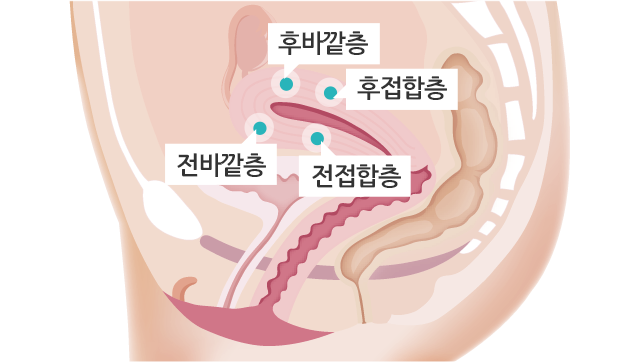
Adenomyosis can be classified into 4 types (sclerotic, nodular, cystic and diffuse), and we tailor the treatment to each type.Adenomyosis varies in size and type from person to person, so before HIFU it needs an accurate treatment plan and precise technique that does not affect surrounding tissue.
-
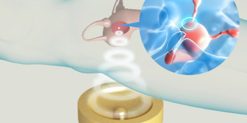 Precise high-intensity ultrasound targeting, down to the cell level
Preserving the uterine lining and surrounding tissue, focused ultrasound precisely treats · destroys the lesion down to the cell level.
-
 Only the adenomyosis tissue is gradually destroyed, with no damage to normal tissue
Without incision or bleeding, only the fibroid or adenomyosis tissue is precisely targeted and destroyed for a more complete treatment.
-
Assessment and treatment
Before treatment, we check whether a lesion could become malignant and look for hidden malignant lesions.

-
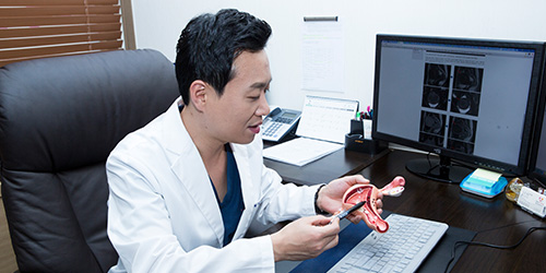
Treatment review and aftercare plan
After treatment, we explain the results and review them objectively
and make an aftercare plan.
-
Regular check-up monitoring
We combine ultrasound and MRI images
to monitor your progress after treatment.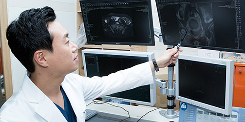
-
Follow-up care
Based on the aftercare plan, we check at set intervals (from the day after treatment, then monthly, quarterly, every six months and yearly) how much the treated lesion has shrunk and whether there is a risk of recurrence.
-
Check-up scheduling
From before HIFU until your regular check-ups after discharge, we take care of every detail.

-
Dedicated 1:1 service
A dedicated coordinator at the clinic looks after
your follow-up visits and recovery.
Consultation menu
Family sites



Awards
Social media

(Seolleung Station Exit 6, Line 2 / Suin-Bundang Line) Tel: +82-2-542-0100 ┃ Business Registration No. 106-91-90905 Representative: Jae-Seong Lee