MRI-Integrated HIFU for Uterine Fibroids and Adenomyosis | Aegis Roen HIFU Center




Main menu

Section menu
Page title
Page content
HIFU SYSTEM Aegis Roan Obstetrics and Gynecology Clinic's high-level treatment method that effectively and safely treats fibroids while preserving the uterus and avoiding general anesthesia and invasive surgery.
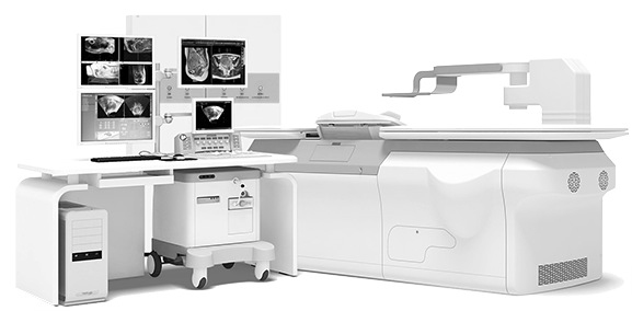

Assessment: Accurately identify the location, size, and shape of tumors (such as uterine fibroids or adenomyosis) using pre-procedure MRI results.


Planning: Establish a treatment plan, including the output size and entry path for the High-Intensity Focused Ultrasound (HIFU) energy.
Execution: Conduct the HIFU treatment guided by the pre-treatment plan, utilizing fused ultrasound-MRI imaging and real-time ultrasound guidance.
3STEP HIFU PROGRAM Since 2010, we have operated a comprehensive MRI-integrated HIFU treatment program focused exclusively on the safe treatment of uterine fibroids and adenomyosis.
MRI scanning to assess the possibility of malignant transformation before the procedure and to analyze hidden malignant lesions.
Collaborative monitoring of ultrasound and MRI images during the procedure.
HIFU treatment briefing and objective evaluation via post-procedure MRI scan.
=
Uterine Fibroid and Adenomyosis Non-Surgical treatment system
MRI scanning analyzes the condition of the uterine fibroids (location, number, size, shape, etc.), the blood supply and blood flow of the fibroids, major adjacent organs and structures, and the feasibility of securing the ultrasound firing path. Furthermore, precise ultrasound (analysis) determines the number of uterine fibroids to be removed, the number of treatment slices and volume of each slice, the fibroid ablation margin, and the correlation between the fibroid and adjacent organs.
-
Analysis of Past Treated Lesions
A detailed investigation is conducted to confirm the extent to which previously treated lesions have been absorbed.
-
Analysis of Recurrence Potential
We check whether there is any evidence of recurrence at the margin of
the treated lesion. -
Analysis of New Lesions
We check whether there is any evidence of new lesions developing in
other areas. -
Analysis of Malignant Transformation
We check whether there is any evidence or possibility of malignant change
Consultation menu
Family sites



Awards
Social media

(Seolleung Station Exit 6, Line 2 / Suin-Bundang Line) Tel: +82-2-542-0100 ┃ Business Registration No. 106-91-90905 Representative: Jae-Seong Lee