How Ultrasound-Guided HIFU Treats Uterine Disease | Aegis Roen HIFU Center




Main menu

Section menu
Page title
Uterine Disease Treatment System
With almost no harm to the body, uterine fibroids · adenomyosis
are treated safely and effectively with our HIFU system
are treated safely and effectively with our HIFU system
Page content
AegisRoen HIFU 3D treatment imaging
Safer HIFU treatment backed by wide clinical experience
HIFU uses high-intensity focused ultrasound to destroy tumor tissue in three dimensions, so it can be adapted to the size, number, shape and location of each tumor
for the best result. The destroyed tissue is gradually absorbed by the body over time.
High-end Artificial Intelligence HIFU
Safer HIFU treatment backed by wide clinical experience
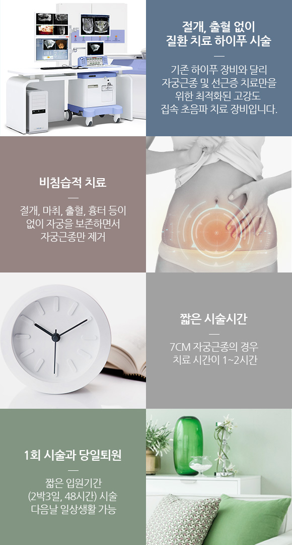
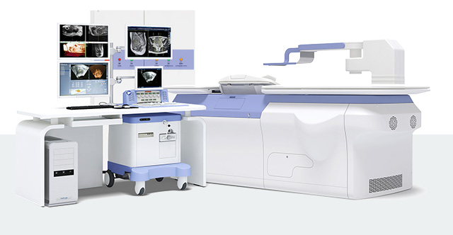
High-end Artificial Intelligence
JC200D uterine disease treatment system It focuses many small points of energy to destroy tumor tissue in three dimensions, so treatment can be tailored to the size, number, shape and location of each tumor. The destroyed tissue is gradually absorbed by the body over time.
JC200D uterine disease treatment system It focuses many small points of energy to destroy tumor tissue in three dimensions, so treatment can be tailored to the size, number, shape and location of each tumor. The destroyed tissue is gradually absorbed by the body over time.
-
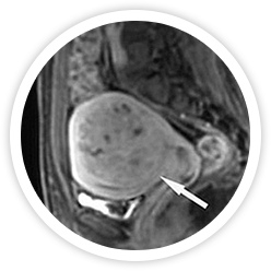 Before treatment
-
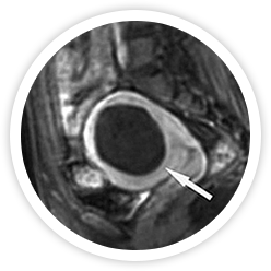 Right after treatment
-
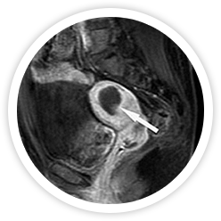 6 months after treatment
-
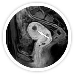 1 year after treatment
-
Thermal Effect
Focused ultrasound heats and
destroys tumor cells -
Cavitations Effect
Rising temperature increases pressure inside the cells,
destroying the tumor cells -
Damage to tumor-nourishing
blood capillariesDestroys the small blood vessels
that feed the tumor, so the tumor cells
die -
Imagery interpretation
Uterine tissue
Precise AI-assisted image analysis system -
Damage Minimization
Minimal damage to surrounding tissue (bowel, bladder, pelvic nerves)
and fast return to normal -
Safety & Availablility
Safety and effectiveness of HIFU officially recognized
(Ministry of Health and Welfare Notice No. 2013-30)
Powerful focused
ultrasound treatment
ultrasound treatment
Highly efficient,
precisely focused treatment
precisely focused treatment
Preserves the endometrium;
destroyed tissue is absorbed
destroyed tissue is absorbed
Advanced technology for thorough treatment
for thorough treatment Artificial Intelligence - Software System
for thorough treatment Artificial Intelligence - Software System
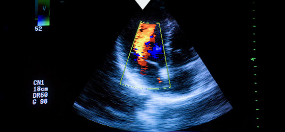
Precise HIFU
with no damage to normal tissue Precise Control
with no damage to normal tissue Precise Control
Specialized check-up system
Check-up process and tests
Artificial Intelligence Check up System For accurate diagnosis of tumors in the uterus, Aegis Roen OB-GYN runs a specialized uterine check-up program to follow every possible change during recovery after HIFU.
Check-up process and tests
Artificial Intelligence Check up System For accurate diagnosis of tumors in the uterus, Aegis Roen OB-GYN runs a specialized uterine check-up program to follow every possible change during recovery after HIFU.
-
 Specialized comprehensive check-up for uterine disease
-
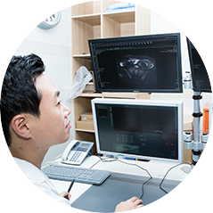 Detailed review of test results
-
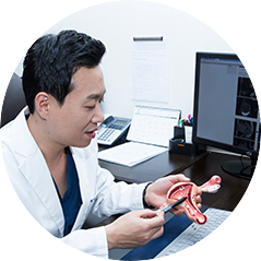 Choosing a treatment
-
Basic tests
Early detection and treatment of fibroids, vaginitis and other conditions

-
Gynecologic uterine exam
In-depth uterine tests for accurate HIFU treatment

-
Women's cancer screening
Early detection and treatment of gynecologic cancers (cervical, ovarian, breast cancer, etc.)
Facilities for check-ups and treatment
New HIFU system and premium ultrasound diagnostic equipment
-
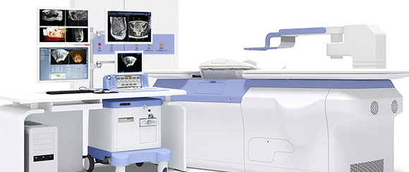 New HIFU system for uterine disease
(HIFU JC-200D and premium system) -
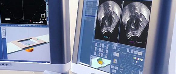 Advanced imaging and diagnostic equipment
(MRI and detailed tests) -
 Designated national cancer screening clinic
(early detection and treatment of women's cancers) -
 MOU with KMI
(comprehensive women's health check-up system)
· Complications such as bleeding, infection, and inflammation may occur after surgery or the procedure.
Furthermore, subjective satisfaction levels may vary among individuals. Therefore, please consult with a specialist.
24-hour phone line
+82-2-542-0100
Weekdays10 a.m. – 9 p.m.
Saturday10 a.m. – 6 p.m.
Consultation menu
Family sites



Awards
Social media

(Seolleung Station Exit 6, Line 2 / Suin-Bundang Line) Tel: +82-2-542-0100 ┃ Business Registration No. 106-91-90905 Representative: Jae-Seong Lee