Early Treatment of Adenomyosis | Aegis Roen HIFU Center




Main menu

Section menu
Page title
you have a lifelong chance
to keep your uterus healthy.
Page content
Adenomyosis When the uterine lining or similar tissue grows outside the uterine cavity
(uterine muscle, ovaries, fallopian tubes, peritoneum, ligaments around the uterus, bladder, vaginal wall, etc.),
it is called endometriosis,
and when it grows inside the uterine muscle, it is called adenomyosis.
It is reported to be most common in women in their 40s and 50s.
and it can also
cause infertility and miscarriage.
When the uterine lining or similar tissue grows outside the uterine cavity (uterine muscle, ovaries, fallopian tubes, peritoneum, ligaments around the uterus, bladder, vaginal wall, etc.), it is called endometriosis, and when it grows inside the uterine muscle, it is called adenomyosis.
up about 20% in the last 5 years

increasing in young unmarried women in their 20s? Statistics show that more young women in their 20s and 30s have uterine conditions such as fibroids, adenomyosis and endometriosis. Rates are rising fast in women in their 20s and 30s. Many are not aware of uterine disease, skip regular check-ups, and dismiss severe period pain or back pain as ordinary PMS, missing the right time for treatment.
| 20s | 30s | 40s | 50s | 60s |
|---|---|---|---|---|
| 9,090 women | 56,630 | 144,489 | 77,568 | 12,315 |
-
Cause 1
Persistently high levels of the female hormone estrogen are involved in its growth and can lead to it.
-
Cause 2
Changes in the uterine muscle tissue can make it similar to lining tissue, leading to adenomyosis.
-
Cause 3
Other factors, such as genetics, hormone imbalance and stress
whose lesions have no clear border, depends on advanced
HIFU skillandprecise analysis.
Unlike fibroids, adenomyosis does not form distinct nodules, so it has no clear border. It is usually about as small as a fibroid, and often occurs together with fibroids.
-
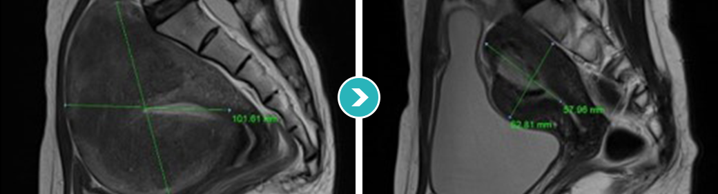 Diffuse type
DIFFUSE TYPE ADENOMYOSIS
Diffuse type
DIFFUSE TYPE ADENOMYOSIS
-
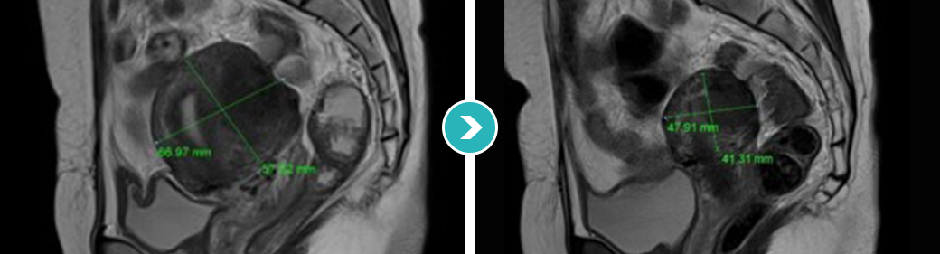 Nodular type
NODULAR TYPE ADENOMYOSIS
Nodular type
NODULAR TYPE ADENOMYOSIS
-
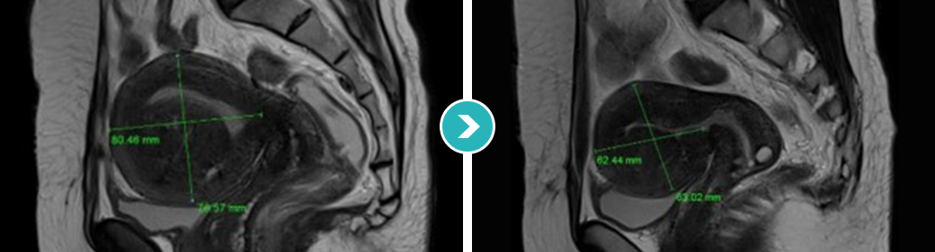 Sclerotic type
SCLEROTIC TYPE ADENOMYOSIS
Sclerotic type
SCLEROTIC TYPE ADENOMYOSIS
-
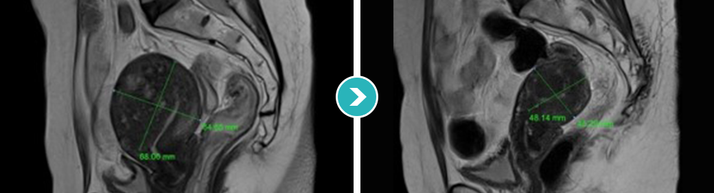 Cystic type
CYSTIC TYPE ADENOMYOSIS
Cystic type
CYSTIC TYPE ADENOMYOSIS
The organs in the body sit close together,
so after HIFU for adenomyosis
the doctor must make sure the lesion was well treated with ultrasound heat
from the inside to just inside its outer edge, and that
the heat did not affect nearby organs or nerves.
This requires a specialist with advanced
HIFU skill.
Future pregnancy and childbirth
may become impossible. If an unmarried woman's gynecologic ultrasound shows fibroids or another uterine condition, she should decide on treatment with a specialist. Treating it with surgery such as removal of the fibroid or the uterus can make future pregnancy and childbirth impossible.
Surgery that removes the whole uterus because the fibroid is large
Surgery that removes the lesion with instruments through small cuts instead of a large abdominal incision
A treatment that uses the heat created when high-intensity ultrasound energy is focused on one point to destroy the tissue
Early adenomyosis treatment system
The Roen AI-assisted HIFU program preserves the uterus, is safer than surgery, keeps pregnancy and childbirth possible, and involves no incision or bleeding
Artificial Intelligence HIFU System
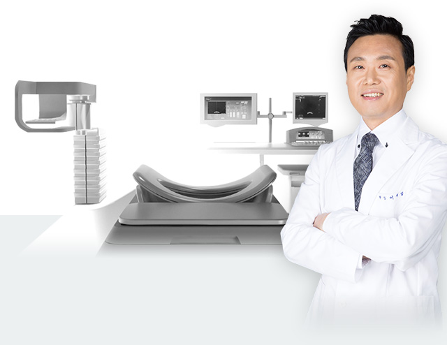
High-resolution MRI images and real-time ultrasound images are combined into a 3D view that tracks the uterus and tumor as they move with the patient's breathing and body movement, so that high-intensity focused ultrasound energy treats them safely.
Artificial Intelligence Check up System
For accurate diagnosis of tumors in the uterus, and to follow every possible change during recovery after HIFU, we run a specialized uterine check-up program for ongoing follow-up.
-
 Non-surgical HIFU to prevent major conditions and
restore normal uterine functionUnlike older HIFU devices, this is a high-intensity focused ultrasound system
High-intensity focused ultrasound -
 Period problems, vaginitis and more
Early diagnosis and treatment of women's conditionsFor unmarried women, gynecologic check-ups and
early treatment are now a must! -
 Comprehensive women's check-up
Personal Woman Total check-up service
-
 Short, simple ∙ safe treatment
Short hospital stay; back to daily life the next day
-
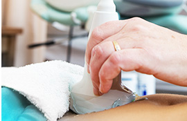 Basic ultrasound
Checks the ovaries, fallopian tubes, uterine lining, uterine body and more for any problems
-
 Cervical cancer screening
Checks for human papillomavirus (HPV) infection
-
 One session, quick discharge
Short hospital stay (1 night, 48 hours)
Back to daily life the next day
Key points
The Roen AI-assisted HIFU program preserves the uterus, is safer than surgery, keeps pregnancy and childbirth possible, and involves no incision or bleeding
and choosing the right treatment for each person is important
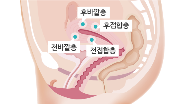
Adenomyosis can be classified into 4 types (sclerotic, nodular, cystic and diffuse), and we tailor the treatment to each type.Adenomyosis varies in size and type from person to person, so before HIFU it needs an accurate treatment plan and precise technique that does not affect surrounding tissue.
-
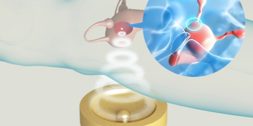 Precise high-intensity ultrasound
targeting, down to the cell levelPreserving the uterine lining and surrounding tissue, focused ultrasound precisely treats · destroys the lesion down to the cell level.
-
 Only the adenomyosis tissue is gradually destroyed,
with no damage to normal tissueWithout incision or bleeding, only the fibroid or adenomyosis tissue is precisely targeted and destroyed for a more complete treatment.
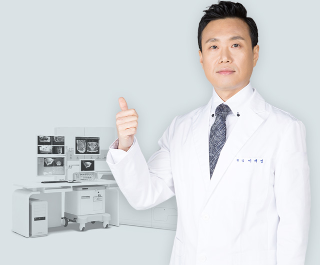
Real Story I first introduced HIFU for uterine fibroids ∙ adenomyosis to achieve successful treatment.
- Former Director, HIFU Center, Department of OB-GYN, Incheon Christian Hospital
- Former HIFU Advisor, Health Insurance Review & Assessment Service (Korea)
- Adjunct Professor, Department of OB-GYN, Inha University College of Medicine
- Adjunct Professor, Incheon St. Mary's Hospital, Catholic University of Korea
- Director, Aegis Roen OB-GYN Clinic
- (formerly Aegis Women's Clinic)
- Director, International HIFU Training Center
- Member, Korean Society of Obstetrics and Gynecology
- Member, Korean Society of Gynecologic Endocrinology
- Member, Korean Society of Gynecologic Endoscopy
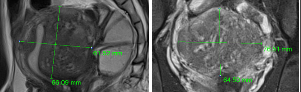
She was diagnosed with adenomyosis because of severe period pain and anal pain, and was admitted for HIFU.
MRI before HIFU showed the uterus enlarged to 6.5 cm, with adenomyosis changes mainly in the back wall.
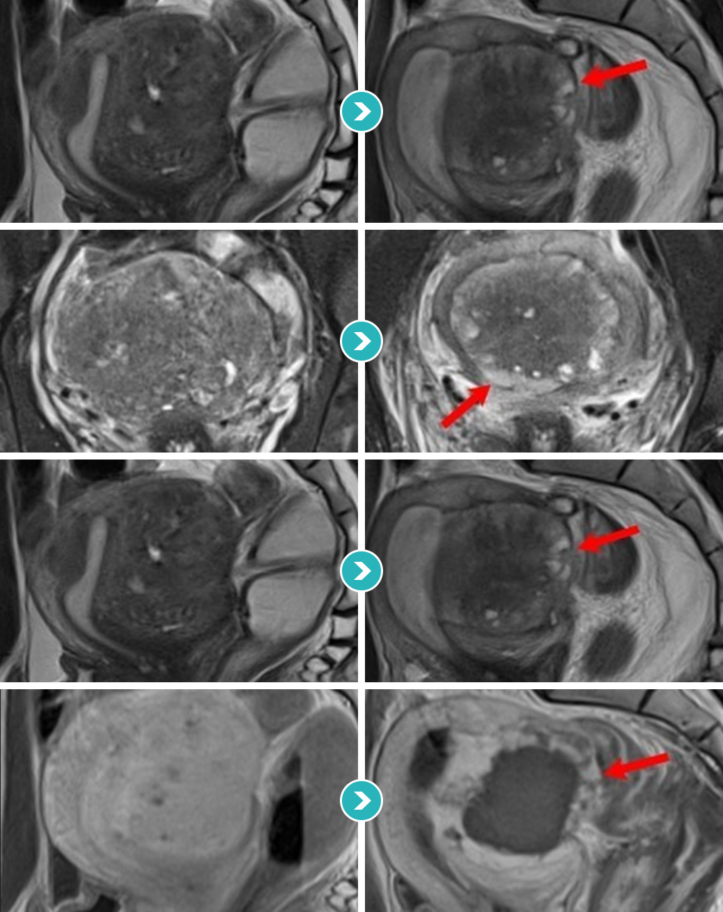
The MRI images below compare before and after HIFU. Over 90% was treated, and the treated area appears gray and black.
A good outcome is expected, and the remaining lesion at the edge is being monitored with hormone treatment.
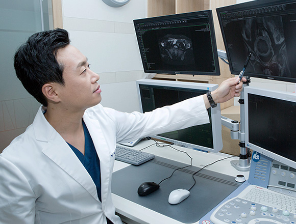
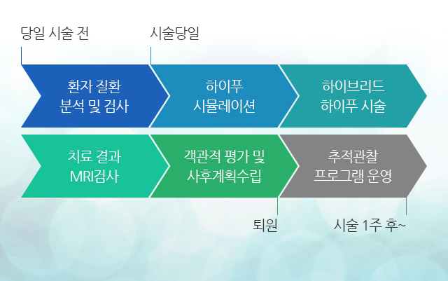
Follow-up Program Personalized aftercare plan = MRI-integrated HIFU at its core

-
Assessment and treatment
Before treatment, we check whether a lesion could become malignant and look for hidden malignant lesions.

-
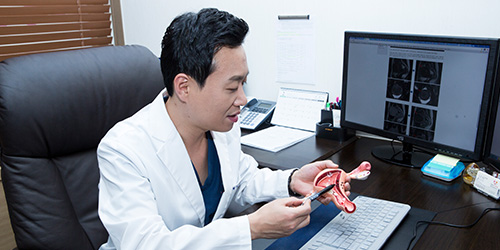
Treatment review and aftercare plan
After treatment, we explain the results and review them objectively
and make an aftercare plan.
-
Regular check-up monitoring
We combine ultrasound and MRI images
to monitor your progress after treatment.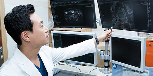
-
Follow-up care
Based on the aftercare plan, we check at set intervals (from the day after treatment, then monthly, quarterly, every six months and yearly) how much the treated lesion has shrunk and whether there is a risk of recurrence.
-
Check-up scheduling
From before HIFU until your regular check-ups after discharge, we take care of every detail.

-
Dedicated 1:1 service
A dedicated coordinator at the clinic looks after
your follow-up visits and recovery.
Consultation menu
Family sites



Awards
Social media

(Seolleung Station Exit 6, Line 2 / Suin-Bundang Line) Tel: +82-2-542-0100 ┃ Business Registration No. 106-91-90905 Representative: Jae-Seong Lee